
Xanthelasma And Xanthomas Pictures: An Eyelid Photo Guide
Key Takeaways
Xanthelasma looks like soft, flat or slightly raised yellow patches on the eyelids, usually near the inner corners. These xanthelasma and xanthomas pictures show how cholesterol deposits around the eyes vary in size, shape and colour, so you can compare them with your own.
- The colour is pale yellow to orange-yellow, clearly different from the skin around it.
- It’s most often on the upper eyelids near the nose, and sometimes on the lower lids too.
- Patches often appear on both eyes, which is very common.
- Xanthomas are similar fatty deposits that can also form elsewhere, such as the elbows, knees or hands.
- Photos help you compare, but only a doctor can confirm what a patch is.
- See real results on our before and after page, or learn about our xanthelasma removal cream.
How To Recognise Xanthelasma In Photos
When you compare your eyelids with the pictures on this page, check these six things. If most of them match, it’s very likely xanthelasma. Our guide to what xanthelasma looks like goes into more detail.
- Colour: pale yellow to orange-yellow, clearly different from the surrounding skin, and not red, brown or white.
- Texture: soft and velvety, flat or slightly raised. It shouldn’t be hard, crusty, scaly or weeping.
- Location: on the eyelids, usually at the inner corners near the nose, and sometimes under the eyes.
- Pattern: often on both eyes, and sometimes on all four eyelids, roughly mirroring each other.
- Size: anything from a spot a few millimetres across to patches covering much of the eyelid.
- Feel: usually painless and not itchy, and it tends to grow slowly over months or years.
Similar deposits elsewhere on the body are usually called xanthomas. A patch that bleeds, hurts, crusts over or changes quickly isn’t typical of xanthelasma, so please show it to your doctor.
Xanthelasma And Xanthoma Photo Gallery
Each photo below has a short note on what to look for. Most show xanthelasma on the eyelids, and a few show other kinds of xanthoma for comparison. Look at the colour, position and texture, and remember that the same condition can look quite different from one person to the next.
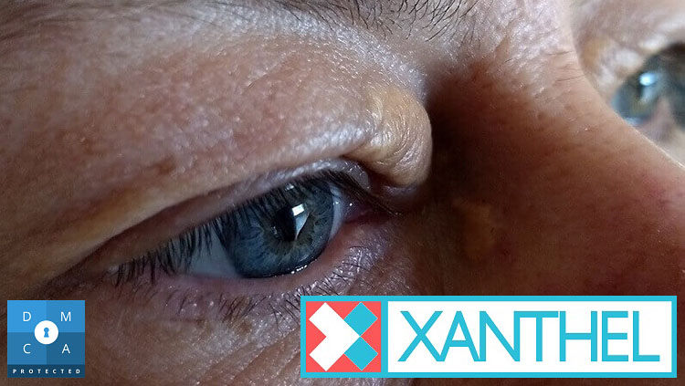
Xanthomatosis Images
Xanthomatosis simply means having several xanthomas. Around the eyes, these fatty deposits usually show up as soft yellow patches on the eyelids.
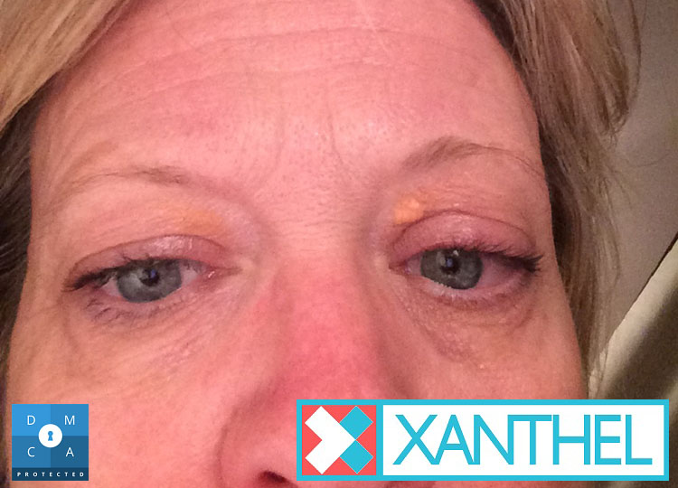
Xanthomas Under The Eye Pictures
Patches aren’t only found on the upper lid. They can form on the lower eyelid too; our guide to cholesterol deposits under the eyes explains more.
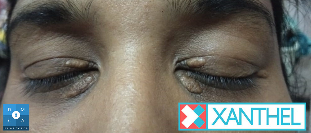
Xanthomas Pictures
Look at the colour first. Xanthomas around the eyes are usually pale yellow to orange-yellow and stand out clearly from the skin around them.
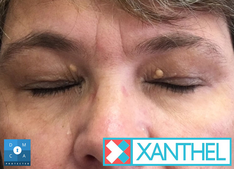
Xanthomas Pictures On The Eyelids
On the eyelids, xanthomas are called xanthelasma. They usually start near the nose and can slowly spread outwards across the lid.
Xanthomas Images
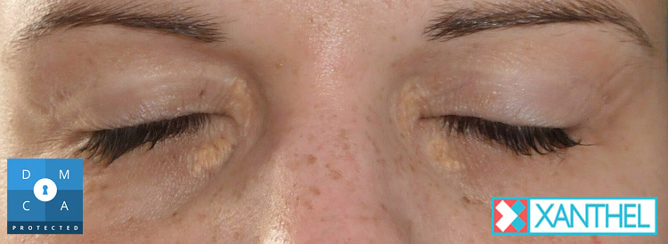
Patches can be flat or slightly raised. Xanthelasma usually feels soft to the touch, not hard, crusty or scaly.
Images Of Xanthomas By The Eye
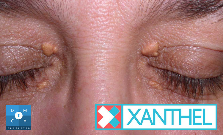
Size varies a lot. Some people have a single spot a few millimetres across, while others have patches covering much of the eyelid.
Xanthoma Pictures
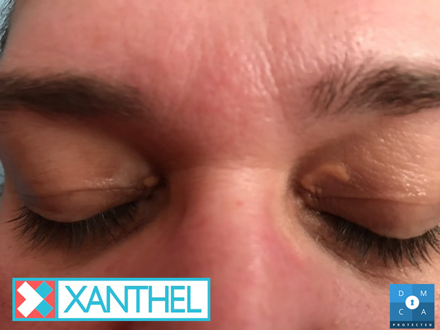
A xanthoma is a build-up of fat-filled cells under the skin. It isn’t contagious, and it isn’t caused by poor hygiene or make-up.
Xanthoma Pictures Of The Eye Area
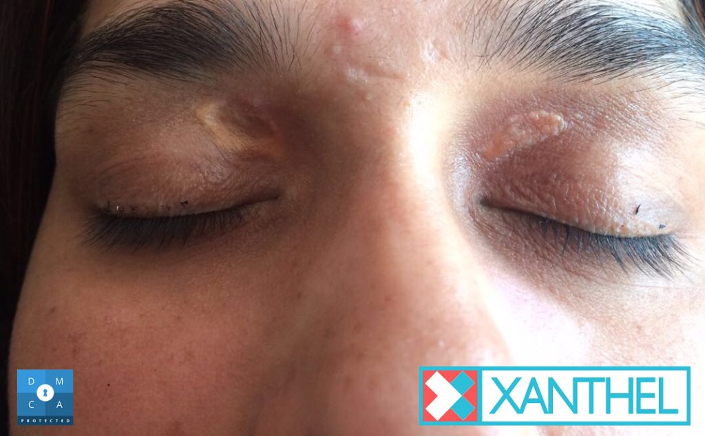
Xanthelasma sits on the skin of the eyelid, not in the eye itself, so it doesn’t usually affect your eyesight.
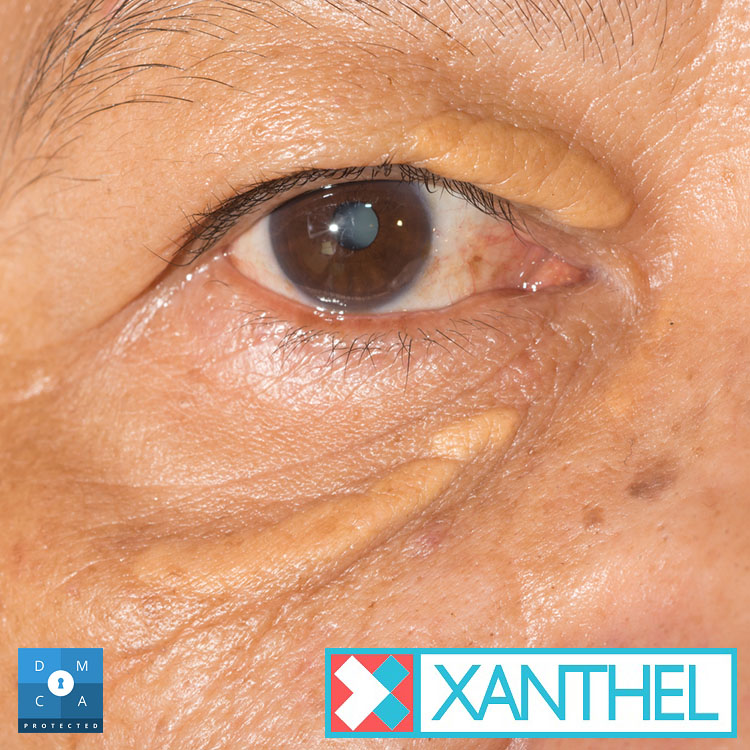
Xanthoma Pictures On The Skin
Xanthomas can also appear away from the eyes, for example on the elbows, knees, hands or along tendons. These are worth showing to a doctor.
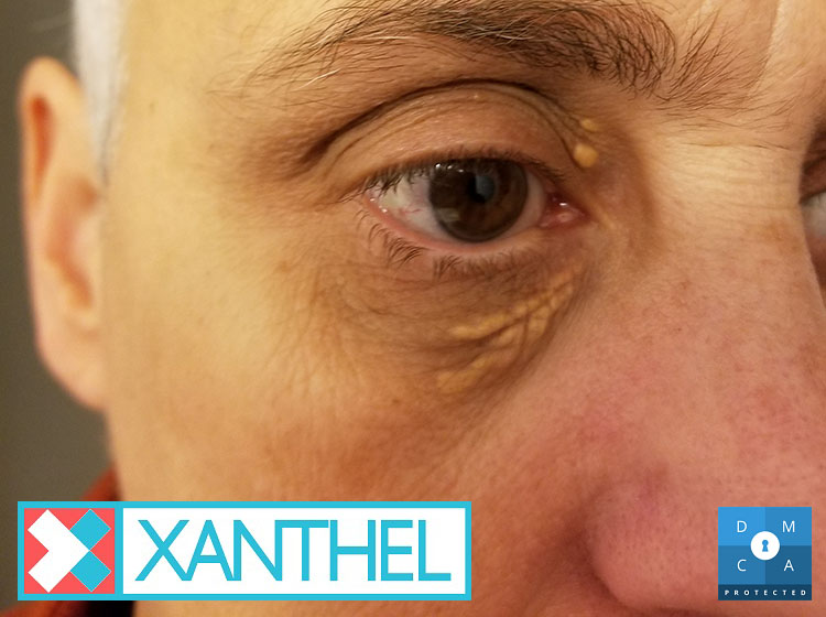
Xanthoma Eye Pictures
Xanthelasma is usually painless and not itchy. If a patch on your eyelid hurts, bleeds or changes quickly, it may be something else.
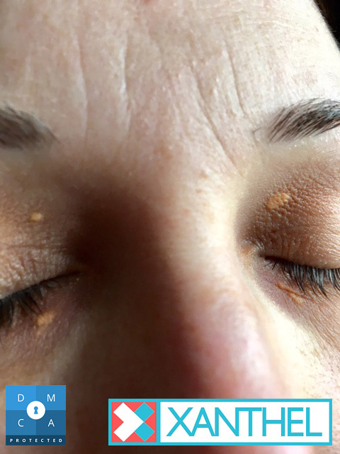
More Xanthoma Images
Patches tend to grow slowly over months or years rather than days. Many people only notice them once they start to spread.
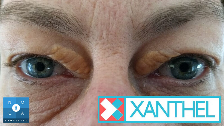
Image Of Xanthelasma: Where It Starts
The upper eyelid, close to the nose, is the most common place for xanthelasma to appear first.
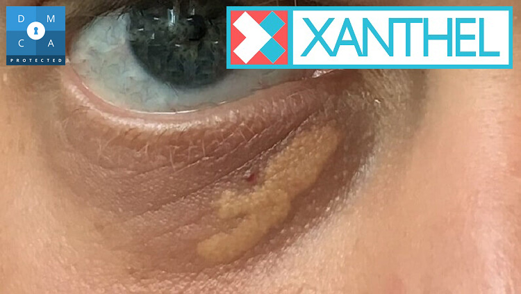
Pictures Of Xanthelasma Of The Eyelid
Xanthelasma palpebrarum is the full medical name. “Palpebrarum” just means “of the eyelids”, which is where these patches almost always appear.
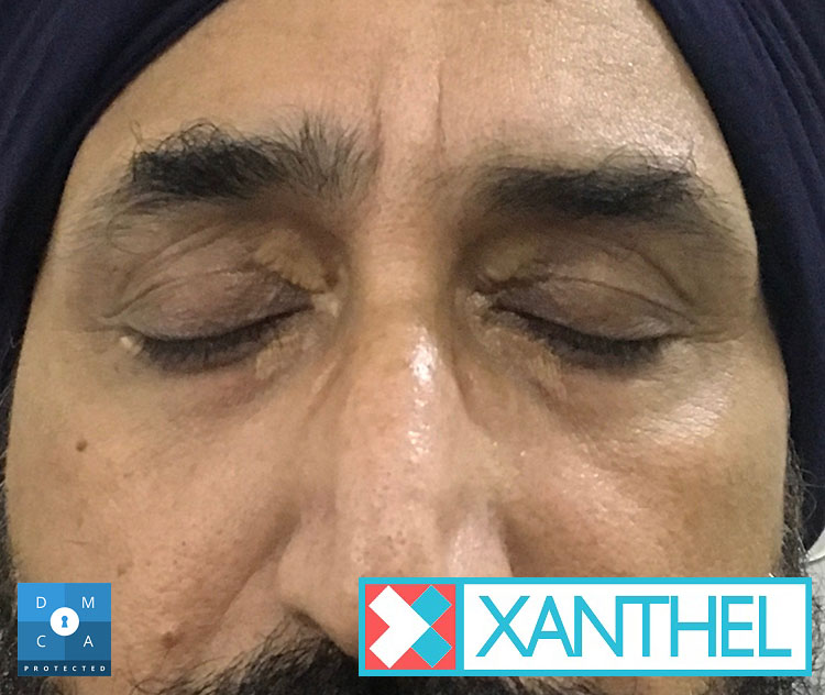
Images Of Xanthelasma: Colour And Shape
Patches are often oval or irregular in shape, with fairly clear edges, and range from pale lemon to a deeper orange-yellow.
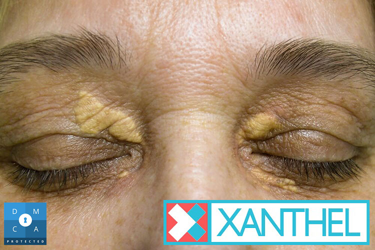
Eyelid Cholesterol Deposit Pictures
Around half of people with xanthelasma have normal cholesterol levels, according to NIH StatPearls, but it’s still worth having a blood test.
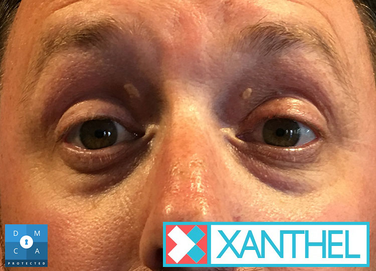
Xanthelasma Of The Eyelid Images
Patches can appear on one eyelid or several. When both eyes are affected it’s called bilateral xanthelasma, which is very common.
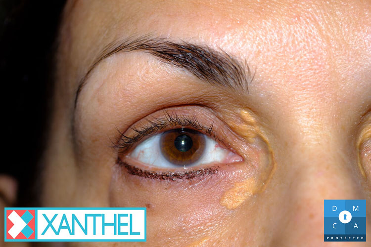
Xanthelasma Image
Xanthelasma is most common in adults over 40 and in women, though it can appear in younger people and in men too.
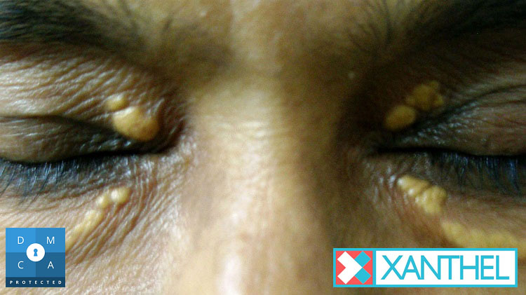
Xanthelasma And Xanthomatosis Pictures
Xanthelasma is the most common type of xanthoma. If you have deposits elsewhere on your body as well, mention them to your doctor.
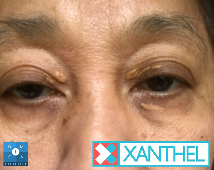
Xanthelasma Or Xanthoma? Pictures Compared
The words are often used interchangeably. Xanthoma is the umbrella term and xanthelasma is the name for xanthomas on the eyelids.
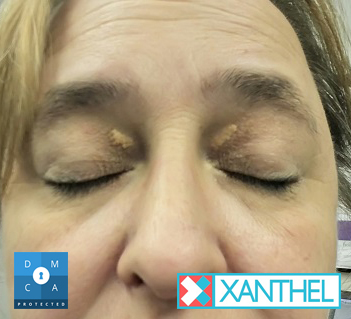
Xanthelasma And Xanthomas Images
Patches don’t usually go away on their own. Once they’ve formed, they tend to stay the same or slowly get bigger.
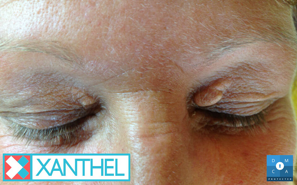
Xanthelasma And Xanthoma Photos
Lighting changes how patches look in photos. Compare yours in natural daylight, without make-up, for the clearest view.

Images Of Xanthelasma
On darker skin tones, xanthelasma can look more orange or tan than yellow, but it has the same soft, flat texture.
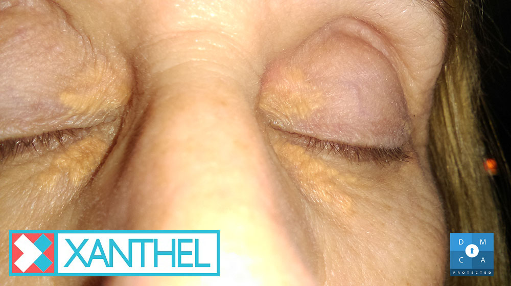
Pictures Of Planar Xanthelasma
Xanthelasma is a flat, or “plane”, xanthoma. That’s why it often looks like a smooth yellow stain rather than a lump.
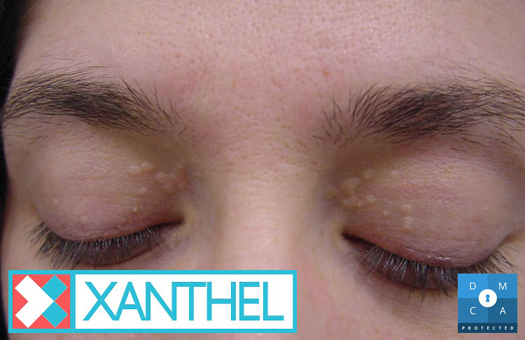
Pictures Of Cholesterol Deposits Around The Eyes
Cholesterol deposits around the eyes can sit above, below or at the inner corner of the eye, and sometimes in all three places.
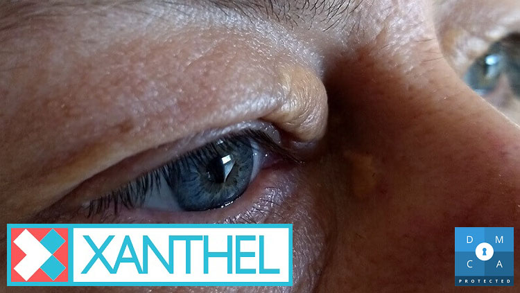
Pictures Of Xanthelasma Around The Eyelids
The skin around the eyes is thin and delicate, which is why harsh home remedies such as acids or garlic can burn it.
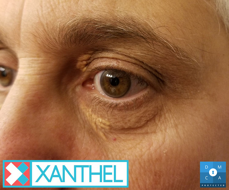
Pictures Of Xanthomas
Xanthomas are linked to how your body handles fats. Raised cholesterol, diabetes and an underactive thyroid are common xanthelasma causes.
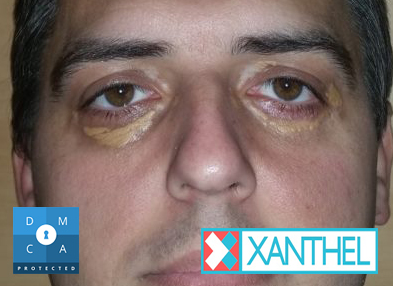
Pictures Of Cholesterol Deposits On The Eyelids
Doctors can usually recognise xanthelasma just by looking at it, so a biopsy is rarely needed. They may suggest a blood test instead.
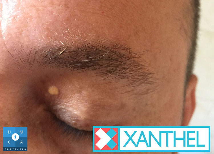
Picture Of Xanthelasma
Xanthelasma isn’t harmful in itself, but because it can be linked to heart health, it’s worth mentioning to your doctor.